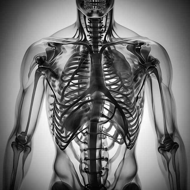
Medical imaging for lung and chest health, including X-rays, CT scans, and MRI, offers critical insights into respiratory conditions. These techniques visualise internal structures, aiding in detecting pneumonia and other infections through identifying inflammation, fluid buildup, and growths. CT scans provide detailed cross-sectional images for accurate diagnoses, while MRI offers a radiation-free alternative for monitoring treatment progress. AI-powered imaging systems further enhance detection capabilities and diagnosis speed.
In today’s digital age, detecting pneumonia and chest infections has evolved significantly through advanced medical imaging. This article delves into the essential tools and techniques used to assess lung health, focusing on technologies like X-rays, CT scans, and emerging methods. We explore how these imaging modalities reveal unseen insights, aiding in accurate diagnosis and effective treatment of conditions affecting the chest. By understanding these methods, healthcare professionals can navigate the complex landscape of lung disease diagnosis, ensuring better patient outcomes.
Medical Imaging for Lung Health: Tools and Techniques
Medical imaging plays a pivotal role in detecting pneumonia and other respiratory infections, offering crucial insights into lung health. Techniques such as X-rays, computed tomography (CT) scans, and magnetic resonance imaging (MRI) are instrumental in visualizing internal structures of the chest. These non-invasive methods enable healthcare professionals to identify signs of inflammation, fluid accumulation, or abnormal growths within the lungs, aiding in accurate diagnosis.
X-rays remain a fundamental tool due to their accessibility and effectiveness in detecting consolidation and opacities indicative of pneumonia. CT scans, with their high resolution, provide detailed cross-sectional images, facilitating the identification of subtle abnormalities and helping differentiate between bacterial and viral infections. MRI offers an even more comprehensive view, especially valuable for assessing underlying lung conditions and tracking treatment progress without exposing patients to radiation.
Detecting Pneumonia: X-rays, CT Scans, and More
Detecting pneumonia involves utilizing various medical imaging techniques targeting the lungs and chest. X-rays remain a standard initial step, offering quick and accessible insights into potential consolidations or opacities indicative of pneumonia. While limited in detail, they serve as a crucial first screen.
For more comprehensive evaluation, computer tomography (CT) scans provide detailed cross-sectional images of the lungs, allowing for better visualization of subtle abnormalities. This advanced medical imaging technique aids in distinguishing pneumonia from other lung conditions, enhancing diagnostic accuracy.
Analyzing Chest Infections: Visualizing the Unseen
In the realm of medical imaging, detecting pneumonia and other chest infections goes beyond what meets the eye. By delving into the intricate world of X-rays, CT scans, and ultrasounds, healthcare professionals can visualize the unseen intricacies of the lungs and chest. These advanced imaging techniques play a pivotal role in identifying subtle anomalies that might not be apparent through physical examination alone.
Through high-resolution images, medical experts can detect inflammation, consolidations, and pleural effusions—all indicative of potential lung infections. For instance, a CT scan can paint a detailed picture of the lung parenchyma, revealing areas of decreased oxygenation or the presence of pneumothorax. This visual representation empowers doctors to make accurate diagnoses, choose appropriate treatment paths, and ultimately, enhance patient outcomes in cases of pneumonia and chest infections.
Advanced Technologies in Lung Disease Diagnosis
The field of medicine has witnessed a significant evolution in detecting and diagnosing lung diseases, particularly pneumonia, thanks to advanced technologies in medical imaging for lung and chest. Computerized Tomography (CT) scans have become a cornerstone in this process, offering high-resolution cross-sectional images that can reveal subtle abnormalities indicative of infections or inflammations within the lungs. These detailed visualizations allow healthcare professionals to make more accurate diagnoses, which is crucial in managing these conditions effectively.
Furthermore, Artificial Intelligence (AI)-powered imaging systems are transforming lung disease diagnosis by enhancing the detection capabilities and providing quantitative measurements. AI algorithms can analyze medical images with remarkable speed and accuracy, identifying patterns associated with various lung pathologies, including pneumonia and its complications. This innovative approach promises improved patient outcomes by enabling early and precise identification of infections, leading to timely interventions.
Medical imaging plays a pivotal role in detecting pneumonia and chest infections, offering valuable insights into lung health. From traditional X-rays and CT scans to cutting-edge technologies, these tools enable accurate visualization of internal structures, aiding in timely diagnosis and effective treatment strategies. By leveraging advanced imaging techniques, healthcare professionals can navigate the complex landscape of lung diseases, ensuring improved patient outcomes and enhanced chest infection management.
