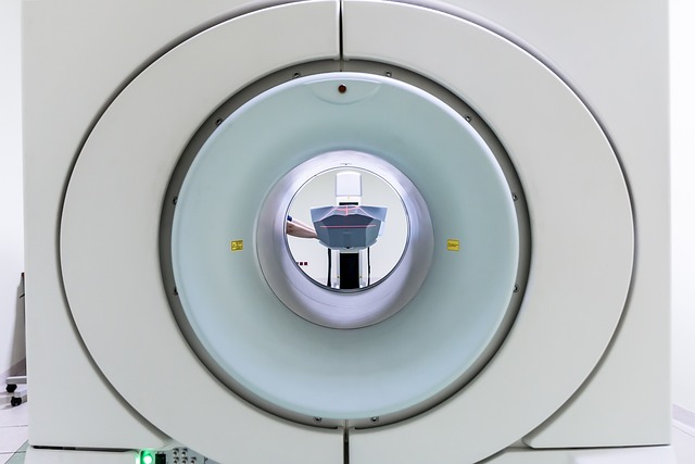
PET scans, along with high-resolution lung CTs, are powerful tools for detecting and diagnosing lung cancer, including metastases. While CTs provide detailed anatomical images, PET scans offer functional information about metabolic activity, helping doctors pinpoint suspicious lesions. Together, these advanced imaging techniques enhance accuracy in identifying metastasis, guiding treatment decisions, and offering a comprehensive understanding of the disease's extent and spread.
“Unveiling the power of Positron Emission Tomography (PET) scans, this article delves into the advanced imaging techniques used to detect and diagnose lung cancer. We explore how PET scans, combined with high-resolution lung CT, offer crucial insights into tumor presence and metastases.
From understanding the fundamentals of PET technology to examining its role in identifying lung cancer and tracking metastases, this guide provides an in-depth look at the modern diagnostic tools transforming patient care.”
Understanding PET Scans: Unraveling the Basics
PET scans, or Positron Emission Tomography, are a powerful tool in detecting and diagnosing lung cancer, including metastases. Unlike traditional imaging methods like high-resolution lung CTs, which provide detailed anatomical images, PET scans offer functional information about the body’s metabolic activity. This distinction is crucial when it comes to identifying cancerous cells, as they often exhibit distinct metabolic patterns compared to healthy tissue.
During a PET scan, a radioactive tracer is introduced into the patient’s bloodstream. This tracer tends to accumulate in areas with heightened metabolic activity, such as tumors. The scanner then detects the energy released by the decaying tracer, allowing healthcare professionals to create detailed images of these metabolic hotspots. In the context of lung cancer, this enables doctors to pinpoint suspicious lesions or masses that may be indicative of both primary tumors and metastases.
High-Resolution Lung CT: Enhancing Visualization
High-resolution lung CT offers a powerful tool for detecting and visualizing lung cancer and its metastases. This advanced imaging technique provides incredibly detailed cross-sectional images of the lungs, allowing healthcare professionals to identify small abnormalities that might be missed by standard X-rays. The enhanced resolution reveals subtle changes in lung tissue, such as nodules, masses, or patterns indicative of cancerous growths and spread.
By producing high-quality pictures, high-resolution lung CT enables precise localization and characterization of lung lesions. This visualization aids in the initial detection, staging, and monitoring of lung cancer, guiding treatment decisions and helping to predict patient outcomes. It’s a significant step forward in early identification and management of this complex disease.
Detecting Lung Cancer: The Role of PET Scans
PET (Positron Emission Tomography) scans play a pivotal role in detecting lung cancer, offering crucial insights that complement traditional high-resolution lung CT imaging. By tracking metabolic activity within the body, PET scans can identify abnormal cell growth and cancerous tumors with remarkable accuracy. This is particularly important in the early stages of lung cancer, when tumors might be too small to visually discern on a CT scan.
The sensitivity of PET scans lies in their ability to detect metastases, which are distant sites of cancer spread. They can visualize areas of increased glucose metabolism, indicative of aggressive tumor growth and potential metastasis. This information is vital for healthcare professionals as it helps in staging the disease, determining treatment options, and predicting patient outcomes. Thus, PET scans provide a comprehensive view, going beyond structural details captured by CT scans, to uncover the biological activity associated with lung cancer.
Identifying Metastases: Advanced PET Imaging Techniques
PET scans, aided by advanced imaging techniques like high-resolution lung CTs, play a pivotal role in identifying metastases associated with lung cancer. These cutting-edge technologies offer a detailed look inside the body, allowing healthcare professionals to detect distant tumors that may have spread from the original lung tumor. By visualizing metabolic activity, PET scans can pinpoint areas of increased cell growth and division, indicative of cancerous cells.
High-resolution lung CTs enhance this process by providing crisp, clear images of the lungs, enabling radiologists to correlate PET scan findings with specific anatomical locations. This integration of data from different imaging modalities improves accuracy in identifying metastases, guiding treatment decisions, and offering a more comprehensive understanding of the cancer’s extent and spread.
PET scans, combined with high-resolution lung CT, offer a powerful tool in detecting both lung cancer and metastases. By providing detailed images of the body’s metabolic activity, PET scans can pinpoint suspicious growths and distant spread, enabling early intervention and improved patient outcomes. Integrating this technology with advanced visualization techniques like high-resolution lung CT ensures comprehensive cancer detection and management.
