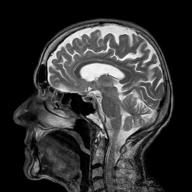
Non-invasive lung CT scans provide high-resolution cross-sectional images of the lungs, enabling accurate diagnosis and personalized treatment for respiratory conditions like COPD, interstitial lung diseases, and pneumonia, while minimizing radiation exposure compared to invasive procedures.
Non-invasive imaging techniques are transforming the diagnosis of respiratory disorders, offering safe, detailed insights into lung health. This article explores cutting-edge approaches, focusing on the high-resolution lung CT scan and advanced technologies beyond traditional X-rays. We delve into their advantages, applications, and how they enhance accurate, efficient diagnoses, ultimately improving patient care and outcomes for diverse respiratory conditions. By understanding these non-invasive methods, healthcare professionals can navigate the evolving landscape of respiratory imaging.
Understanding Non-Invasive Imaging for Respiratory Health
Non-invasive imaging plays a pivotal role in diagnosing and managing respiratory disorders, offering safer alternatives to invasive procedures like lung biopsies. Techniques such as chest X-rays, high-resolution computed tomography (HRCT) scans, and magnetic resonance imaging (MRI) provide detailed visual information about the lungs’ structure and function without requiring penetration of the skin or major internal organs.
Among these, HRCT scans stand out due to their ability to produce high-resolution images of the lung tissues, enabling healthcare providers to detect subtle abnormalities associated with conditions like chronic obstructive pulmonary disease (COPD), interstitial lung diseases, and pneumonia. Unlike a regular chest X-ray, a lung CT scan can capture intricate details of the lung architecture, including tiny airways, blood vessels, and parenchyma, facilitating more accurate diagnoses and guiding personalized treatment plans.
High-Resolution Lung CT Scan: Advantages and Applications
High-Resolution Lung CT (HRCT) scans offer a non-invasive way to visualize detailed images of the lungs, providing unique advantages in diagnosing and monitoring respiratory disorders. This advanced imaging technique captures high-resolution cross-sectional images, allowing radiologists to identify subtle abnormalities that may be missed by standard chest X-rays.
The primary applications of HRCT include evaluating pneumothorax, bronchiolitis, interstitial lung diseases, and detecting early signs of chronic obstructive pulmonary disease (COPD) or tuberculosis. By providing clear, detailed images, HRCT enables accurate diagnosis, guiding treatment plans, and tracking disease progression over time, making it a valuable tool in respiratory medicine.
Advanced Techniques: Beyond Traditional X-rays
In recent years, non-invasive imaging techniques have advanced dramatically, offering improved diagnostic capabilities for respiratory disorders beyond traditional X-rays. One such technique is the high-resolution lung CT (HRCT) scan. This advanced method provides detailed cross-sectional images of the lungs, allowing healthcare professionals to detect subtle abnormalities that may be missed by standard chest X-rays. HRCT scans can visualize airway patterns, parenchymal structures, and blood vessels with remarkable clarity, aiding in the early detection and diagnosis of conditions such as chronic obstructive pulmonary disease (COPD), interstitial lung diseases, and pneumonias.
Additionally, other non-invasive methods like magnetic resonance imaging (MRI) and ultrasound are gaining traction for their ability to offer comprehensive views of the respiratory system without exposure to ionizing radiation. MRI scans, for instance, use strong magnetic fields and radio waves to produce detailed images, while ultrasound utilizes high-frequency sound waves to visualize internal structures in real time. These cutting-edge techniques not only enhance diagnostic precision but also play a crucial role in monitoring disease progression and evaluating treatment responses, thereby improving overall patient care.
Safety and Efficiency in Respiratory Disorder Diagnosis
Non-invasive imaging techniques have revolutionized the diagnosis of respiratory disorders, offering safe and efficient alternatives to traditional methods. One such advanced tool is the lung CT scan, which provides high-resolution cross-sectional images of the lungs, allowing healthcare professionals to detect abnormalities with remarkable accuracy. This technology minimizes radiation exposure, making it a safer option compared to other diagnostic procedures.
Compared to invasive techniques, lung CT scans offer significant advantages in terms of comfort and speed. Patients undergo a quick and relatively painless scan, providing immediate visual feedback for doctors to make informed decisions. The efficiency of this method enables early detection and management of respiratory conditions, potentially improving patient outcomes and quality of life.
Non-invasive imaging techniques, particularly high-resolution lung CT scans and advanced technologies, have revolutionized the diagnosis of respiratory disorders. By offering detailed insights into lung structures without the risks associated with invasive procedures, these methods enable early detection and precise planning for treatment. Incorporating such innovations in healthcare not only enhances patient safety but also improves outcomes for those suffering from respiratory conditions. In light of their effectiveness and growing accessibility, lung CT scans and advanced non-invasive imaging will continue to be indispensable tools in the global effort to combat respiratory diseases.
