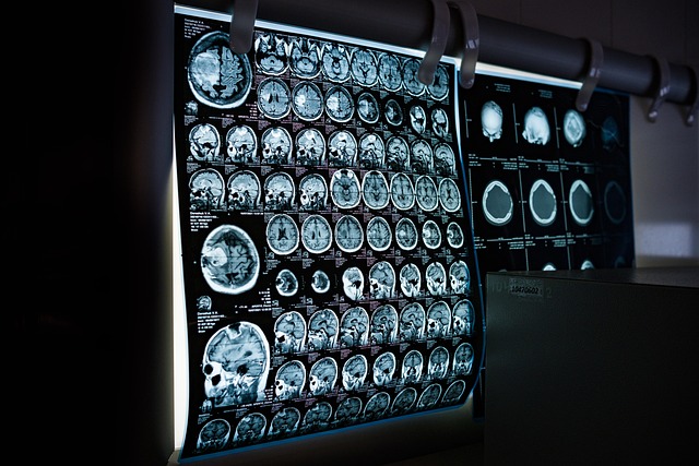
High-Resolution Lung CT (HRCT) is a powerful non-invasive imaging tool that enhances diagnosis and treatment of lung conditions by visualizing ventilation and perfusion patterns. HRCT's advanced resolution allows radiologists to detect subtle abnormalities in air spaces, consolidation, and emphysema, crucial for conditions like COPD, interstitial lung diseases, and pulmonary embolisms. By combining nuclear medicine tracers with HRCT, V/Q scans offer detailed insights into lung function, enabling precise treatment strategies and improved patient outcomes.
In the realm of medical imaging, understanding lung ventilation and perfusion is key to diagnosing pulmonary conditions. This article explores the critical role of nuclear medicine in V/Q scans, highlighting its applications and benefits in clinical practice. We begin by demystifying lung ventilation and perfusion fundamentals, before delving into how high-resolution lung CT enhances image quality. Subsequently, we discuss nuclear medicine’s unique contribution to V/Q scans, shedding light on its invaluable role in precision diagnostics.
Understanding Lung Ventilation and Perfusion: The Basics
Lung ventilation and perfusion, often abbreviated as V/Q, refer to the process by which air reaches (ventilation) and blood flows through (perfusion) the lungs. This crucial pair of functions ensures optimal oxygen exchange in the respiratory system. Ventilation involves the movement of air in and out of the lungs, while perfusion is the circulation of blood within the lung’s capillaries, facilitating the transfer of oxygen into the bloodstream and carbon dioxide out.
High-resolution lung CT (HRCT) plays a pivotal role in visualizing these processes. By providing detailed images of the lung structure, HRCT enables healthcare professionals to assess ventilation and perfusion patterns accurately. This non-invasive technique is particularly valuable for diagnosing conditions like chronic obstructive pulmonary disease (COPD), interstitial lung diseases, and pulmonary embolisms, where abnormalities in V/Q relationships can offer vital clues for diagnosis and treatment planning.
High-Resolution Lung CT: Enhancing Image Quality
High-Resolution Lung CT (HRCT) has significantly enhanced the image quality of lung ventilation and perfusion scans, including V/Q scans. By providing incredibly detailed cross-sectional images of the lungs, HRCT allows radiologists to detect even subtle abnormalities that might be missed by lower-resolution techniques. This advanced imaging modality employs a series of thin slices, offering a more comprehensive view of lung parenchyma, bronchioles, and vascular structures.
The improved resolution enables better assessment of lung diseases, such as interstitial lung diseases, pneumonias, and chronic obstructive pulmonary disease (COPD). HRCT can visualize air spaces, identify areas of consolidation, and quantify the extent of emphysema, all of which are crucial for accurate diagnosis and treatment planning. This technology revolutionizes V/Q scans by providing more precise information, ultimately leading to better patient outcomes.
Nuclear Medicine's Contribution to V/Q Scans
Nuclear medicine plays a pivotal role in providing detailed insights into lung ventilation and perfusion, as evidenced by V/Q scans. This advanced imaging modality offers high-resolution lung CT, enabling radiologists to visualize minute structural changes and abnormalities within the lungs. By tracing the movement of radioisotope tracers, such as technetium or xenon, nuclear medicine provides crucial information about air distribution (ventilation) and blood flow (perfusion).
The fusion of nuclear medicine with computed tomography enhances diagnostic accuracy, allowing for the detection of subtle disparities in lung function. This is particularly beneficial in evaluating conditions like pulmonary embolism, interstitial lung diseases, or assessing the effectiveness of ventilation strategies in critical care settings. High-resolution lung CT scans from nuclear medicine studies offer a comprehensive view, helping healthcare professionals make informed decisions and tailor treatment plans accordingly.
Applications and Benefits in Clinical Practice
Nuclear medicine plays a vital role in assessing lung ventilation and perfusion, with V/Q scans (or lung scintigraphy) being a key application. This non-invasive technique uses radioactive tracers to visualize air distribution and blood flow within the lungs. By combining this with high-resolution lung CT (HRLCT), healthcare professionals gain a detailed understanding of both structural abnormalities and functional impairments.
The benefits in clinical practice are significant, enabling accurate diagnosis and monitoring of conditions such as pulmonary embolism, chronic obstructive pulmonary disease (COPD), and interstitial lung diseases. HRLCT-guided V/Q scans provide critical insights into the extent and pattern of ventilation/perfusion mismatches, guiding personalized treatment plans and improving patient outcomes.
Nuclear medicine plays a vital role in lung imaging, particularly with ventilation and perfusion (V/Q) scans. By combining functional data with anatomical insights from high-resolution lung CT, these advanced techniques enable accurate assessment of lung health. The integration of nuclear medicine with CT enhances diagnostic accuracy, guiding treatment plans for various pulmonary conditions. This comprehensive approach, utilizing high-resolution lung CT and V/Q scanning, is a game-changer in clinical practice, offering better patient outcomes and improved management of respiratory diseases.